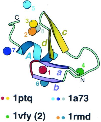
Figure 3.

Metal-binding sites in treble clef fingers. All 42 treble clef finger structures (Fig. 2) are superimposed and metal ions bound at different sites in the structural motif (having zinc ligands from different sites in sequence alignment) are displayed. Metal ions are shown as balls and are numbered from 1 to 8. Protein ribbon corresponds to Cys2 activator-binding domain of protein kinase Cδ (1ptq, residues 241–280). Color coding and labeling of secondary structural elements correspond to Figure 1. PDB codes of representative structures that cover all distinct zinc-binding sites are shown below and colored dots indicate the sites that are present in the structures.
