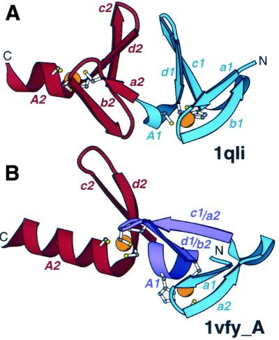
Figure 4.

Arrangement of treble clef pairs. (A) A pair of tandem treble clef motifs in the LIM domain of cysteine-rich protein CRIP (1qli, residues 117–145). (B) Two zinc-binding motifs of the FYVE domain of vps27p protein (1vfy, chain A, residues 173–235). One treble clef finger is colored in blue, the other is colored in red. Secondary structural elements are named according to Figure 1. Indices 1 and 2 refer to the first and second treble clef finger, respectively. The segment that belongs to both treble clef fingers is colored in purple. Side chains of zinc ligands are shown in ball-and-stick representation. Zn2+ is shown as an orange ball.
