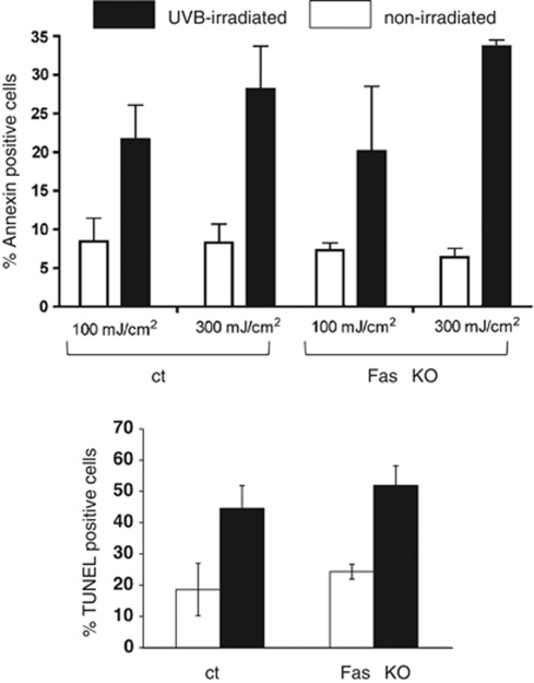
Figure 5.
UVB-induced apoptosis of Fas-negative keratinocytes in vitro. Primary murine keratinocytes in passages 2–4 isolated from control mice (ct) and FasE-KO mice (Fas KO) were irradiated with 100 or 300 mJ/cm2 UVB, respectively. Upper part: FACS analysis of Annexin V exposure on the cell surface was carried out after 16 h. Bar graphs show the percentage of Annexin-V-positive keratinocytes in the propidium-iodide-negative population±S.D. Results are from three control and Fas KO primary isolates analyzed in at least two independent experiments. Lower part: Primary keratinocytes were irradiated with 300 mJ/cm2 UVB and labeled with the TUNEL technique 16 h later. Bar graphs show the percentage of TUNEL-positive keratinocytes in the total adherent cell population±S.D. in two independent experiments using keratinocytes from three control and three FasE-KO mice

