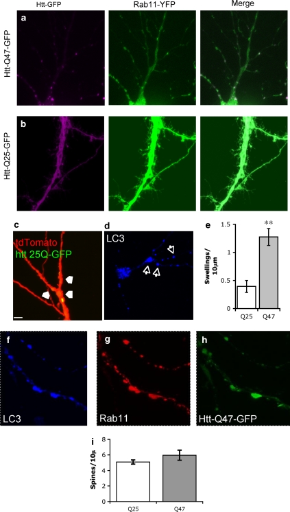
Figure 4.
Altered Rab11 distribution and autophagosomal markers are associated with HttQ47 aggregates in dystrophic dendrites. Neurons were co-transfected with Rab11-YFP and either HttQ47-GFP (a) or HttQ25-GFP (b). Spectral unmixing was used to resolve the YFP and GFP signals. Co-localization of Rab11 and HttQ47 aggregates is seen as bright puncta in the dendrites. (c) Dystrophic swelling (arrows) in HttQ47-GFP cells co-transfected with tdTomato: these swellings were also associated with LC3 accumulation (d). (e) Numbers of dystrophic swellings were quantified in HttQ47-GFP- and HttQ25-GFP-expressing neurons. Data are mean±S.E.M. of 15 determinations; **Significantly different, P<0.01). Neurons triply transfected with Htt- LC3-mRFP (f), Rab11-YFP (g) and HttQ47-GFP (h) were imaged and spectrally unmixed to resolve the GFP/YFP signals and subsequently re-imaged to visualise the LC3-mRFP. The images show the LC3, Rab11 and HttQ47 signals associate in the same dendritic areas. (i) Numbers of spines within 10 μm of an aggregate in HttQ47-GFP-expressing neurons co-transfected with Rab11 compared with numbers in HttQ25-GFP showed no difference

