Figure 1.
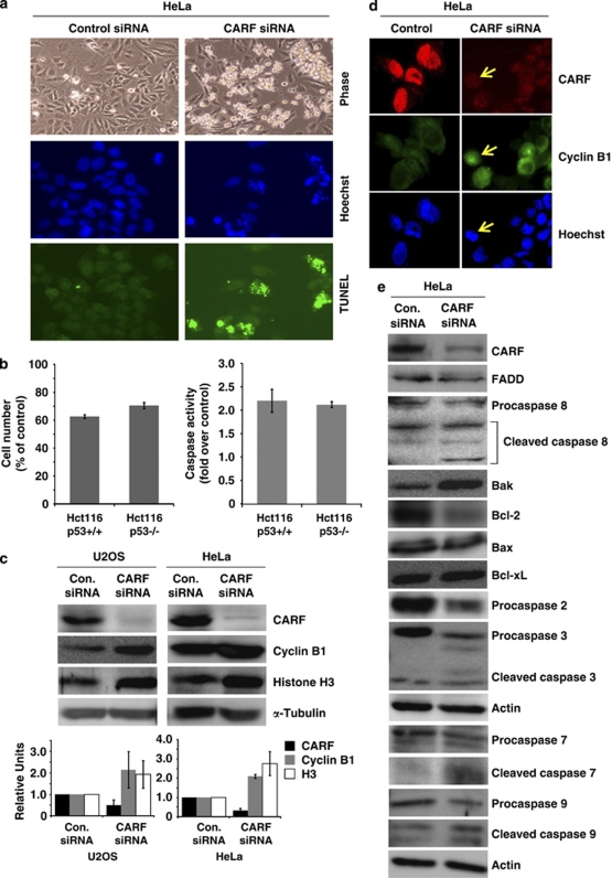
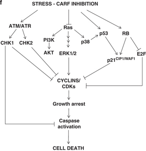
Cell death induced by CARF suppression occurs after mitotic arrest through the mitochondrial stress and caspase-dependent pathway. TUNEL staining of HeLa cells transfected with CARF-targeting siRNA shows increased cell death following CARF suppression (a). Syngeneic p53 +/+ and p53 −/− HCT116 cells showed comparable apoptosis (decreased cell viability, left and increase in caspase 3 activity, right) after CARF suppression (b). CARF-compromised cells underwent mitotic arrest as evidenced by accumulation of cyclin B1 and histone H3 with densitometric quantitation of representative blots from at least three experiments, in which the CARF-suppressed group is shown as fold change over control, which was set as 1 (c). Immunofluorescence shows that cells with reduced CARF levels had increased cyclin B1 nuclear accumulation, as shown by the arrows (d). CARF-compromised HeLa cells were subjected to immunoblotting analyses for proteins constituting the mitochondrial stress and caspase pathways (e). Actin and α-Tubulin were used as loading controls. Graphs are represented as average mean±S.D. (f) Schematic diagram of the hypothetical pathways that lead to cell death following CARF inhibition. We considered that apoptosis induced by CARF suppression might activate multiple pathways, including the ATM/ATR/CHK1/CHK2, Ras-MAPK and/or RB/E2F1 networks


