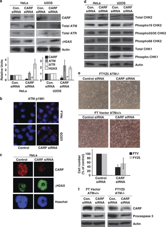
Figure 4.
CARF suppression activates the ATM pathway, but it is not required for cell death. Total ATM, total ATR and γH2AX, as well as CARF levels were analyzed by immunoblotting with densitometric quantitation of representative blots from at least three experiments, in which the CARF-suppressed group is shown as fold change over control, which was set as 1 (a). Phosphorylated ATM at serine 1981 (b) and γH2AX (c) were detected by immunofluorescent staining, wherein, blue stain denotes nuclei. The phosphorylated forms of CHK2, including phosphorylation at serine 19, serines 33/35 and threonine 68, total CHK1 and phosphorylated CHK1 were examined by western blotting (d). ATM +/+ and null cells were transfected with CARF siRNA, in which apoptosis is shown as rounded, floating cells in the images (e), and cleavage of procaspase 3 was detected by immunoblotting (f). Actin was used as loading control and Hoechst 33 258 was used for nuclear staining. Graphs are represented as average mean±S.D.

