Figure 2.
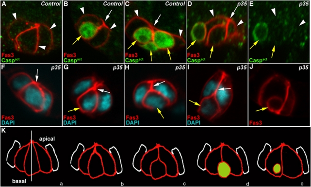
Supernumerary PCs die by apoptosis in a stereotyped manner. Confocal images of control upd-Gal4/+ follicles (A–C) and upd-Gal4/+ UAS-p35/+ follicles in which PCs express the baculovirus caspase inhibitor, p35 (D–J). The apical side of PCs is always to the top. In all images, except (E), the morphology of PCs is revealed by immunostaining specifically for membrane Fas3 (red). Immunodetection of activated caspases (green), which confirms the identity of dying polar cells, was carried out in (A–E). (D) and (E) are views of the same group of PCs. DAPI staining was used to reveal nuclei in (F–I). (F–J) represent a series of different groups of three PCs, which illustrate what may be the temporal progression of morphological changes occurring during PC elimination, in particular, initial apical constriction (F and G, white arrows), followed by apical detachment (H and I, white arrows) as the supernumerary PC rounds up, reduces in size and takes a basal position (H–J, yellow arrows). (K) Model for supernumerary PC elimination. PCs are in red and adjacent follicle cells in white. (a–e) Represent successive steps during elimination of one cell in a group of three PCs. (a) Group of equivalent cells. The white vertical line indicates the axis of radial symmetry around which the group of equivalent cells is organized. The shrinking supernumerary PC progressively loses its apical contact (b–d) and becomes spherical (c–e). (c–e) The green color indicates the timing of caspase activation detection in the dying cell

