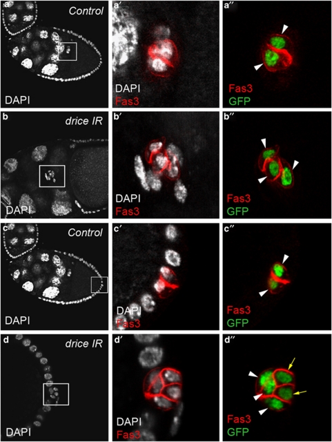
Figure 3.
Drice is implicated in PC apoptosis. Confocal images of control upd-Gal4/+ UAS-H2B:YFP/+ (a and c) and upd-Gal4/+ UAS-driceIR/+ UAS-H2B:YFP/+ (b and d) stage 10 ovarian follicles stained with DAPI (white), Fas3 antibodies (red) and GFP antibodies (green) that, respectively, label all nuclei, PC membranes and PC nuclei. Anterior is to the left, as well as the apical side of PCs in (a, c and d). The PCs and surrounding border cells are migrating between nurse cells in (b). (a′–d′) and (a″–d″) are magnified views of the boxed areas in the corresponding a–d panels. Note that in the control, pairs of PCs are present at a given extremity of stage 10 follicle (a″ and c″, white arrowheads), while expression of a drice RNAi transgene leads to the presence of more than two PCs at both anterior and posterior poles at the same stage (b″ and d″, white arrowheads and yellow arrows to indicate the supernumerary PCs that have begun to shrink and shift basally). Also note that upon expression of a drice RNAi transgene, excess anterior PCs surrounded by border cells are delayed in their migration toward the oocyte at stage 10 (b and b′) compared with control PC pairs that have already reached the oocyte at the same stage (a and a′)

