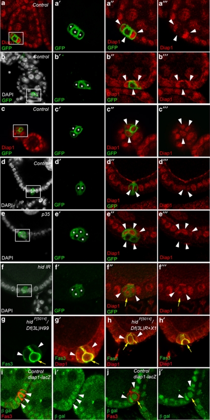
Figure 7.
Hid controls Diap1 downregulation in PCs destined to die. Confocal images of ovarian follicles stained with DAPI to label nuclei (white), GFP antibodies to label PC membranes (green) and Diap1 antibodies (red) (a–f) or with Fas3 antibodies to label PC membranes (green) and Diap1 antibodies (red) (g and h) or with Fas3 antibodies to label PC membranes (red) and β-galactosidase antibodies (green) to detect diap1-lacZ enhancer trap expression (i and j). The apical side of the follicular epithelium in which the PCs are embedded is toward the top. (a′–f′), (a″–f″) and (a′″–f′″) are magnified views of the boxed areas in the corresponding a–f panels. In (a′–f′), each PC is indicated by an asterisk. In (a″–e″), (a′″–e′″) and (i and i′), it is not possible to distinguish between PC destined to survive or die, so all PCs are indicated by white arrowheads. In (f″, f′″, g, g′, h, h′, j and j′), white arrowheads and yellow arrows mark PCs destined to survive or die, respectively, as indicated by the round shape, smaller size and more basal position, indicating progressive exclusion of the latter from the group. (a–d) Control: upd-Gal4/+ UAS-mCD8:GFP/+, (e) upd-Gal4/+ UAS-mCD8:GFP/UAS-p35, (f) upd-Gal4/+ UAS-mCD8:GFP/+ UAS-hid-IR/+, (g and h) hidP(5014)/ Df(3L)H99 (g) and hidP(5014)/Df(3L)R+X1 (h) escapers and (i and j) yw; thj5C8/TM3Sb ovaries

