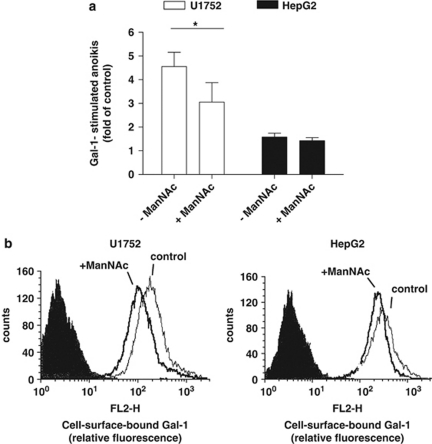
Figure 5.
ManNAc supplementation reduces Gal-1 stimulated cell death and Gal-1 binding. (a) Gal-1-mediated cell death in U1752 and HepG2 cells previously cultured for 48 h with 5 mM or without ManNAc supplementation. Cells were then subjected to suspension culture with continued ManNAc supplementation and Gal-1 was added as indicated. Anoikis rates were determined after 24 h on the basis of the pre-G1 fraction from DNA-histograms. Anoikis is expressed as fold of the respective controls in the absence of Gal-1. Data represent mean±S.E.M. of at least three independent experiments (*P<0.05, one tailed t-test). (b) Binding of biotinylated Gal-1 to U1752 (left panel) or to HepG2 (right panel) cells that had been cultured for 48 h in the presence of ManNAc (+ManNAc) or vehicle (control). Flow cytometry analysis shows control (light), ManNAc-treated (bold) cells and background fluorescence via binding of indocarbocyanine-streptavidin conjugate in the absence of biotinylated Gal-1 (shaded)

