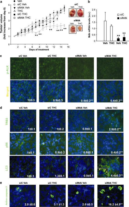
Figure 7.
In vivo silencing of Mdk sensitizes T98 cell-derived tumors to THC antitumoral action. (a) Effect of Mdk silencing and THC (15 mg/kg per day) treatment on the growth of tumor xenografts derived from T98 cells (mean±S.D.; n=5 for each condition, **P<0.01 from the rest of the treatments). siRNAs were administered at days 1 and 7 of vehicle or THC treatment. Representative photographs of siC and siMdk, vehicle and THC-treated tumors are shown. (b) Mdk mRNA levels in T98 cell-derived tumor xenografts as determined by quantitative real-time PCR. Data correspond to Mdk mRNA levels relative to one of the siC vehicle-treated tumors (mean±S.D.; n=5 for each condition; **P<0.01 from vehicle and THC-treated, siC-transfected tumors). (c) Effect of Mdk silencing and THC treatment on phospho-ALK immunostaing of T98 cell-derived tumor xenografts. Values inside the photomicrograhs are expressed as the phospho-ALK-stained area relative to the number of nuclei in each field and correspond to 10 fields of three different tumors for each condition. Data are normalized using siC-transfected vehicle-treated tumors as a reference. Representative photomicrographs are shown (mean±S.D.; **P<0.01 from vehicle-treated, siC-transfected tumors). (d) Effect of Mdk silencing and THC treatment on TRB3 (upper panel), phospho-S6 (middle panel) or LC3 (lower panel) immunostaining of T98 cell-derived tumor xenografts. Values inside the photomicrographs are expressed as the TRB3-, phospho-S6- or LC3-stained area relative to the number of nuclei in each field and correspond to 10 fields of three different tumors for each condition. Data are normalized using siC-transfected vehicle-treated tumors as a reference. Representative photomicrographs are shown (mean±S.D.; **P<0.01 from vehicle-treated, siC-transfected tumors). Inset: high-magnification photomicrograph of the arrow-pointed cell. Arrows within the inset point to LC3 dots. (e) Effect of THC on apoptosis of T98 cell-derived tumor xenografts. Values inside the photomicrographs are expressed as the percentage of active caspase-3 positive cells relative to the total number of nuclei in each field and correspond to 10 representative fields of three different tumors for each condition. Representative photomicrographs are shown (mean±S.D.; **P<0.01 from vehicle-treated, siC-transfected tumors)

