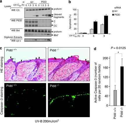
Figure 6.
PIDD deficiency sensitizes cells to UV-C induced apoptosis. (a and b) Hacat cells were transfected with the indicated siRNA and subjected to different times of 50 J/m2 UV-C irradiation. (a) Markers of apoptosis, Bid and caspase-3 cleavage as well as Cytochrome c release were analysed by western-blotting. See also Supplementary Figure S5A. (b) Apoptosis was quantified by Hoechst staining and show the mean of two independent experiments. (c and d) PIDD+/+ or −/− mice were irradiated as described in ‘experimental procedures'. Apoptosis was detected by HE staining (arrowheads indicate sunburn cells) and active caspase-3 staining (c) and quantification (d). See also Supplementary Figure S5b. Data shown are representative of at least three independent experiments and reported as mean ±S.D. P-value for statistical significance was calculated using the Student's t-test and considered significant (*)

