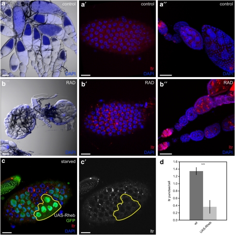
Figure 3.
IIS/TOR signaling controls autophagy in Drosophila ovaries. (a–b″) Injection of RAD leads to small ovaries lacking vitellogenic stages (b) and a strong accumulation of autophagolysosomes in FCs (b′) and GCs (b″). Control ovaries are of normal size (a) and barely show LTR staining in FCs (a′) or GCs (a″). (c and c′) Generation of stage 7 FC clones overexpressing Rheb using the flp-out-Gal4/UAS method results in cells with high (strong GFP signal) and low (weak GFP signal) transgene expression. Only cells with bright GFP signals and enlarged nuclei (as an indication of enhanced cell size due to Rheb overexpression) were considered for the analyses. (d) Quantification of LTR staining in Rheb overexpressing clones compared with WT cells. Error bars show S.D. of the mean, n=8, ***P<0.001. Scale bars: (a and b) 100 μm, (a′, b′, c and c′) 10 μm, (a″ and b″) 50 μm. Genotypes: (a–b) y w, (c) hs flp/+act4CD24Gal4 UAS-GFP/UAS-RhebEP50.084

