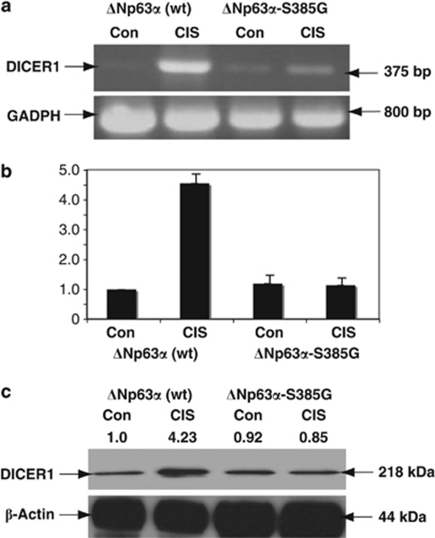
Figure 1.
p-ΔNp63α upregulates DICER1 expression upon CIS exposure. Wild-type ΔNp63α cells (p63wt) or ΔNp63α-S385G cells (p63mut) were exposed to Con (−) or 10 μg/ml CIS (+) for 24 h. Total RNA and protein were isolated and analyzed for DICER1 expression. (a) PCR assay. (b) qPCR assay of DICER1 expression. Data were normalized against GAPDH levels and plotted as relative units (RU), with measurements obtained from wild-type ΔNp63α cells treated with Con set as 1. Experiments were performed in triplicate with±S.D. as indicated (P<0.01). (c) Immunoblotting with anti-DICER1 antibodies (levels of DICER1 were quantified and normalized against β-actin protein level, and the values obtained from untreated wild-type ΔNp63α cells are designated as (1)). As loading controls, we used the GAPDH mRNA levels for PCR and the β-actin protein levels for immunoblotting

