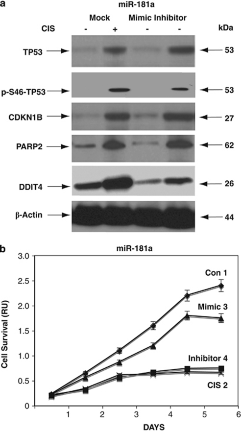
Figure 6.
CIS-induced modulation of miR-181a expression leads to the activation of cell cycle arrest and apoptotic markers. Wild-type ΔNp63α cells were transfected with control (mock), mimic, or inhibitor for miR-181a for 24 h and then exposed to Con (−) or 10 μg/ml CIS (+) for an additional 24 h. (a) Protein levels of TP53, p-TP53, PARP2, CDKN1B, and DDIT4 were examined by immunoblotting with the indicated antibodies and loading levels were tested with an anti-β-actin antibody. (b) CIS-induced modulation of cell survival by miR-181a. Wild-type ΔNp63α cells were transfected with control (mock, curves 1 and 2), miR-181a mimic (curve 3), or miR-181a inhibitor (curve 4) and then exposed to Con (curves 1, 3, and 4) or 10 μg/ml CIS (curve 2) for 0–120 h. Cell survival was assessed at 24, 48, 72, 96, and 120 h by MTT cell proliferation assay. Absorbance readings were taken using a SpectraMax M2e Microplate fluorescence reader (Molecular Devices) at 570 and 650 nm wavelengths. All samples were run in triplicate. Experiments were performed in triplicate with ±S.D. as indicated (<0.05)

