Figure 1.
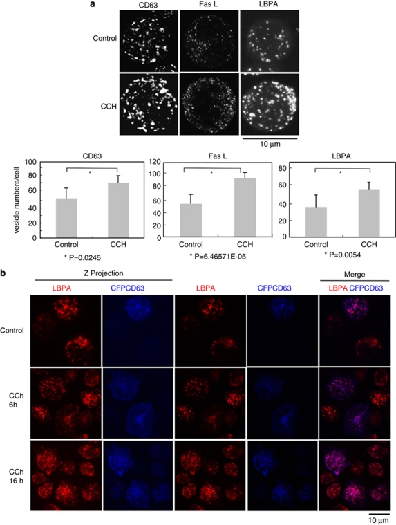
Cellular stimulation induces the formation of mature MVBs. (a) Upper panel, J-HM1-2.2 cells were stimulated with CCh for 6 h and then were imaged by confocal microscopy using antibodies specific for CD63, FasL and LBPA. Lamp-1 is mostly present on the limiting membrane of MVBs, whereas CD63, and particularly LBPA, are abundant in the ILVs,27 and therefore label mature MVBs. LBPA is a phospholipid that participates in the maturation of MVBs33 and constitutes a bonafide marker for ILVs of mature MVBs.33 The z-axis projections for each antigen corresponding to representative cells from three independent experiments are represented. Lower panel shows quantitative analysis of vesicles. Vesicle numbers were recorded from at least 20 cells per group, chosen randomly as indicated in Materials and Methods. Results represent average number of vesicles/cell±S.D. of three independent experiments. (b) J-HM1-2.2 cells, transfected with CFP-CD63, were stimulated with CCh (for 6 and 16 h), and mature MVBs were visualised with anti-LBPA antibody (red) as indicated in Materials and Methods. Cells were imaged by confocal microscopy and representative (n=3 independent experiments), single optical sections (0.4 μm thick) and merged images (coincident labelling appearing pink) are shown in the right side. In the left side, z-axis projection images of LBPA and CFP-CD63 are shown

