Figure 5.
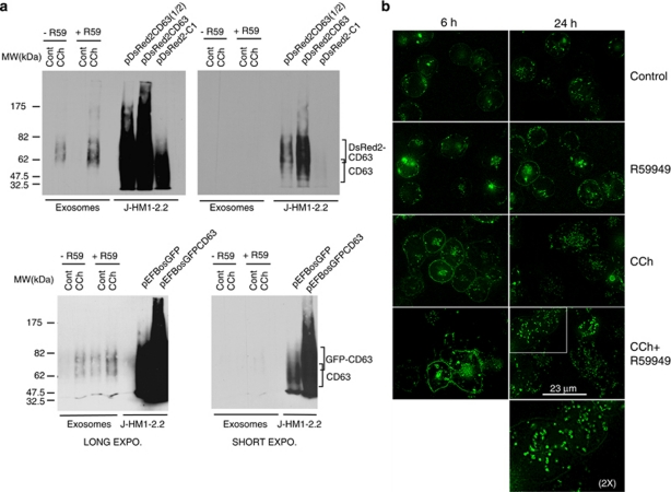
Expression of the reporters for MVBs/exosomes in living cells. (a) J-HM1-2.2 cells expressing DsRed2-CD63 (upper panels) or GFP-CD63 (lower panels) were stimulated with CCh for 10 h in the presence or the absence of R59949 inhibitor (R59, 10 μM), and the isolated exosomes were analysed by WB with anti-CD63 to detect the chimerical CD63 molecules. Different exposures of the same blot are shown to visualise both the chimeras and the endogenous CD63. In the right-side lanes of each blot, lysates from cells expressing or not expressing the CD63 chimeras were run as a reference. (b) Cells expressing GFP-CD63 were stimulated or not expressing with CCh for 6 and 24 h in the presence or the absence of R59949 to visualise the formation and fate of MVBs in living cells. The CCh- and R59949-induced increase in GFP-CD63 at the plasma membrane was probably because of the fusion of the limiting membrane of GFP-CD63+ MVBs (see also Supplementary Videos 3 and 4). The epifluorescence images were improved by image deconvolution as indicated in Materials and Methods, and are representative of the results obtained of more than 50 cells recorded per treatment in four independent experiments. The inset shows a × 2 digital zoom of the indicated area

