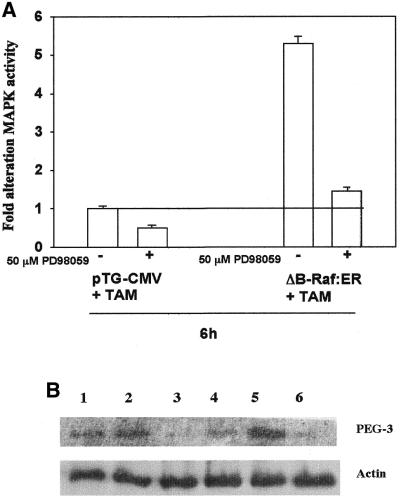
Figure 4.
Specific activation of MAPK by ΔB-Raf:ER increases expression of PEG-3. B1TD cells were infected with either a control recombinant adenovirus (CMV) or a recombinant adenovirus to express ΔB-Raf:ER. Twenty-four hours after infection, cells were treated with 200 nM 4-hydroxytamoxifen (TAM) and either vehicle control (VEH, DMSO) or 50 µM PD98059. Six hours after treatment, cells were washed with PBS and frozen. (A) Cells were lysed and prepared for immune complex kinase assays to determine MAPK activity, as described in Materials and Methods. Data are the means of three separate experiments ± SEM. (B) Cells were lysed in preparation for SDS–PAGE, followed by immunoblotting to determine PEG-3 protein expression levels. A representative experiment is shown n = 3. Lane 1, Ad.vec; lane 2, Ad.vec + TAM; lane 3, Ad.vec + TAM + PD98059; lane 4, Ad.ΔB-Raf:ER; lane 5, Ad.ΔB-Raf:ER + TAM; lane 6, Ad.ΔB-Raf:ER + TAM + PD98059.

