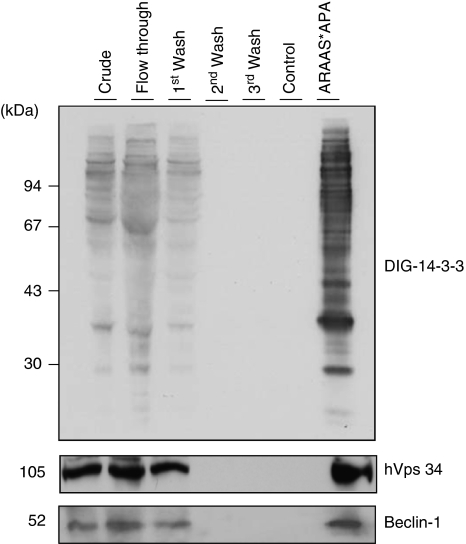
Figure 3.
14-3-3 Affinity chromatography of human HeLa cell extracts. Clarified HeLa cell extract was chromatographed on 14-3-3-Sepharose, as described in Material and Methods. Column fractions were run on SDS/PAGE using 10% Tris-glycine gels, and transferred to nitrocellulose membranes. Amounts of protein run on SDS/PAGE were as follows: extract, flow through and beginning of salt wash (first wash), 40 μg of each; middle and end of salt wash (second wash and third wash, respectively), protein undetectable; control (phospho) peptide pool,<1 μg; and ARAApSAPA (ARAAS*APA) elution pool, 2 μg. Membranes were checked for binding to DIG-14-3-3 (top panel: Published in author's previous work.47 Copyright owners belong to author). Western blots were probed with antibodies against the indicated proteins related to autophagy (bottom panels)

