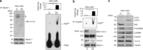
Figure 4.
hVps34 dissociates from 14-3-3 and is activated during C2-ceramide-induced autophagy. HeLa cells continuously growing in serum were left untreated or stimulated with C2-ceramide (10 μM) for 24 h to promote autophagy. (a) Extracts (2 mg) from untreated (U) or C2-ceramide (C2) stimulated HeLa cells were incubated with 1 μg of Beclin-1 (Santa Cruz Biotecnology, Inc.) antibody bound to protein-G-Sepharose. The washed immunoprecipitates were resolved using SDS-PAGE, transferred to nitrocellulose and probed with DIG-14-3-3 (14-3-3 overlay), Beclin-1 (BD Biosciences) and hVps34 (Zymed) antibodies. The asterisk (*) shows an unknown protein band. The arrow corresponds to hVps34 size on DIG-14-3-3. The washed immunoprecipitates were also used for hVps34 in vitro kinase assay (right panel). Autoradiography shows levels of PI3P32. (Quantification of hVps34 in vitro kinase assay expressed as PI(3) fold change, N=3, *P=0.0007, Student t-test.) (b) Extracts (1 mg) from untreated (U) or C2-ceramide (C2) stimulated HeLa cells were incubated with 1 μg of hVps34 (Echelon Biosciences) antibody bound to protein-G-Sepharose. The washed immunoprecipitates were used for hVps34 in vitro kinase assay and also to analyze interaction with 14-3-3 (DIG-14-3-3) and presence of Beclin-1, hVps34 and 14-3-3 proteins in the immunoprecipitates (quantification of hVps34 in vitro kinase assay, N=3, *P=0.0007, Student t-test). Piece of the DIG-14-3-3 shown correspond to hVps34 size. (c) Extracts (30 μg) from HeLa cells continuously growing in serum or stimulated with C2-ceramide (10 μM) for 24 h to promote autophagy were run on SDS-PAGE, transferred to nitrocellulose membranes and probed with indicated antibodies

