
Figure 5.
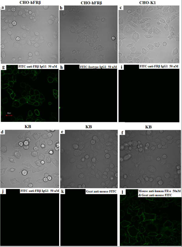
Confocal laser microscopy images show the specific binding of FITC-m909 to CHO-FRβ cells. Human FRβ stably transfected CHO-FRβ cells (a,b,g,h), CHO-K1 cells (c,i), and KB nasopharyngeal epidermoid cells (d-f,j-l) were incubated with 50 nM m909 IgG-FITC for 1 hour at 37°C and were washed three times with cold phosphate-buffered saline. Images (a-f) were captured with transmitted light. Images (g-l) were captured using a charge-coupled device camera with identical settings below the saturation limits. Isotype IgG1-FITC did not give any binding signal in CHO-FRβ cells (b,f). Mouse anti-human FRα mAb together with goat anti-mouse IgG-FITC secondary antibody showed the expression of human FRα on KB cells (f,l). FITC, fluorescein isothiocyanate; FR, folate receptor.