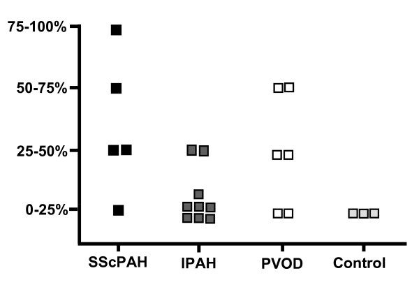
Figure 5.
Amount of phosphorylated plateled-derived growth factor receptor (pPDGFR)-β-positively immunostained cells in intima of the small vessels in SScPAH, IPAH, PVOD and controls. With a cut off of 25% cell staining, a trend was shown (P = 0.09) in favor of more positive cell immunoreactivity in small vasculature in SScPAH patients vs. IPAH patients. Each image represents one case.

