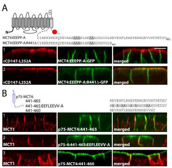
Figure 5. Identification of an additional BLSS within MCT4.
(A) To confirm the BLSS activity of the glutamate and proline residues they were mutated in MCT4-GFP and MCT4:R441Δ-GFP and constructs were stably expressed in MDCK-rCD147-L252A cells. Polarized cells were fixed and immunolabeled with anti-rat CD147 antibody (red). (B) Polarized MDCK cells stably expressing p75-MCT4:441-465 and mutated and truncated p75-MCT4:441-465 constructs were fixed and immunolabeled with anti-p75 antibody (green) to detect the transgenes and anti-MCT4 antibody (red) to detect endogenous MCT4 (red). Confocal images are shown as X-Z images of representative sections of from the Z-stack. Bar=10μm, all images are same scale.

