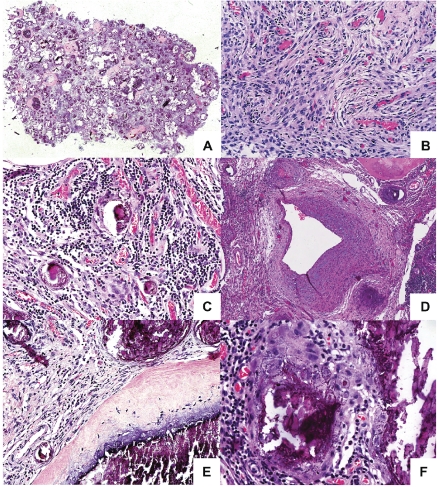
Figure 2.
A heterogeneous tumor with areas of calcification and bone formation. (A) Tumor seen at low power showing cellular areas with areas of calcification and ossification. (B) The cellular areas are composed of plump, elongated cells with meningothelial features. (C) Prominent lymphoplasmacytic infiltrate is present within the cellular areas. (D) The highly vascularized tumour also contain dysplastic blood vessels. (E) The areas of calcification show crystalline deposits of calcium merge seamlessly with woven bone. No osteoblastic rimming is seen. (F) An interlacing chicken-wire-like calcification is seen in several areas.

