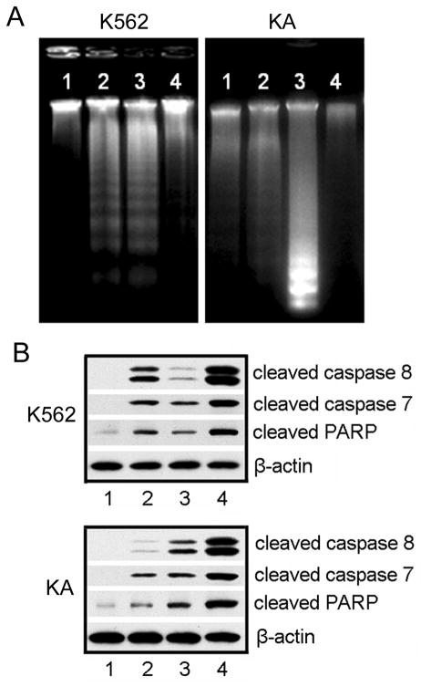
Figure 3.
A. DNA fragmentation in Leukemia cells after different treatments. Genomic DNA was isolated from K562 or KA cells. DNA ladders were visualized under UV light with ethidium bromide staining. K562 cells (a) were treated with: 1, no treatment; 2, 1.99×10⁻⁶ mol/L DNR; 3, 0.58 mg/L Fe3O4 nanoparticles with 1.99×10⁻⁶ mol/L DNR; 4, 0.58 mg/L Fe3O4 nanoparticles. KA cells (b) were treated with: 1, 0.58 mg/L Fe3O4 nanoparticles; 2, 1.99×10⁻⁶ mol/L DNR; 3, 0.58 mg/L Fe3O4 nanoparticles with 1.99×10⁻⁶ mol/L DNR; 4, no treatment. B. Western blotting analysis of activated caspases after various treatments. K562 and KA cell lysates were prepared from the cells without treatment (1), treated with 1.99×10⁻⁶ mol/L DNR (2), 0.58 mg/L Fe3O4 nanoparticles (3), or 0.58 mg/L Fe3O4 nanoparticles and 1.99×10⁻⁶ mol/L DNR (4). The following antibodies were used: anti-cleaved Caspase-8, anti-cleaved Caspase-7, and anti-PARP antibody. β-Actin was used as a loading control.

