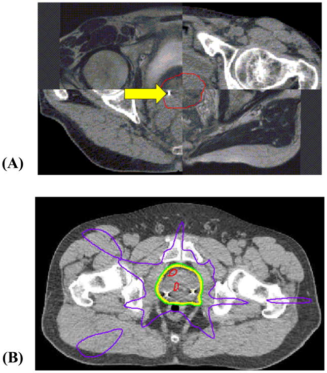
Figure 1.
(A) Checkerboard axial image of the fused MR and CT scans, and the contoured prostate, illustrated for one patient. The final contoured prostate (CTV) is shown in red. A fiducial beacon placed at the right base of the prostate is seen on this CT slice (arrow). (B) Isodose distribution of a 5-field IMRT prostate plan, showing the 50% (purple), 95% (green), 100% (yellow), and 105% (red) isodose lines.

