
Figure 2.
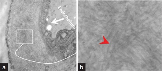
The anterior fat pad aspirate showed amyloid (small box) in the wall of small blood vessels (a). The non-branching random amyloid fi brils with 8.6 nμ diameter were consistent with amyloid (red arrowhead) (b). [Epoxy embedded Glutaraldehyde fi xed section, stained with uranyl acetate and lead citrate. Ultrastructure]