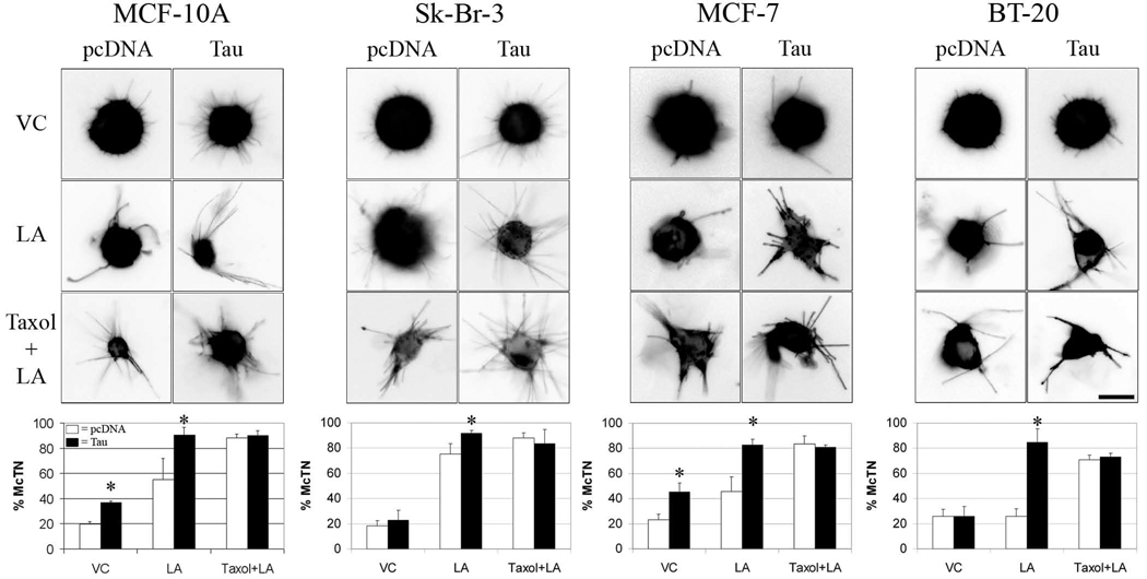
Figure 2. Exogenous tau overexpression stimulates McTN formation.
McTNs were evaluated blindly in live MCF-10A, Sk-Br-3, MCF-7, and BT-20 cells exogenously overexpressing pcDNA (left micrograph panels, white bars) or tau (right micrograph panels, black bars) in VC, LA, and Taxol+LA. Tau overexpression caused distinct morphological alterations in length, thickness and ridigity of McTNs formed compared to pcDNA. Tau-induced McTNs became longer and thicker in VC conditions compared to pcDNA. Treatment of tau-transfected cells with LA increased the length, thickness, and rigidity of McTNs comparable to empty vector cells treated with Taxol+LA. Frequency of McTN formation (lower graphs) revealed that tau significantly increased McTN formation. Data (n=3) are represented as the mean ± s.d. *P<0.05, scale bars = 10µm.

