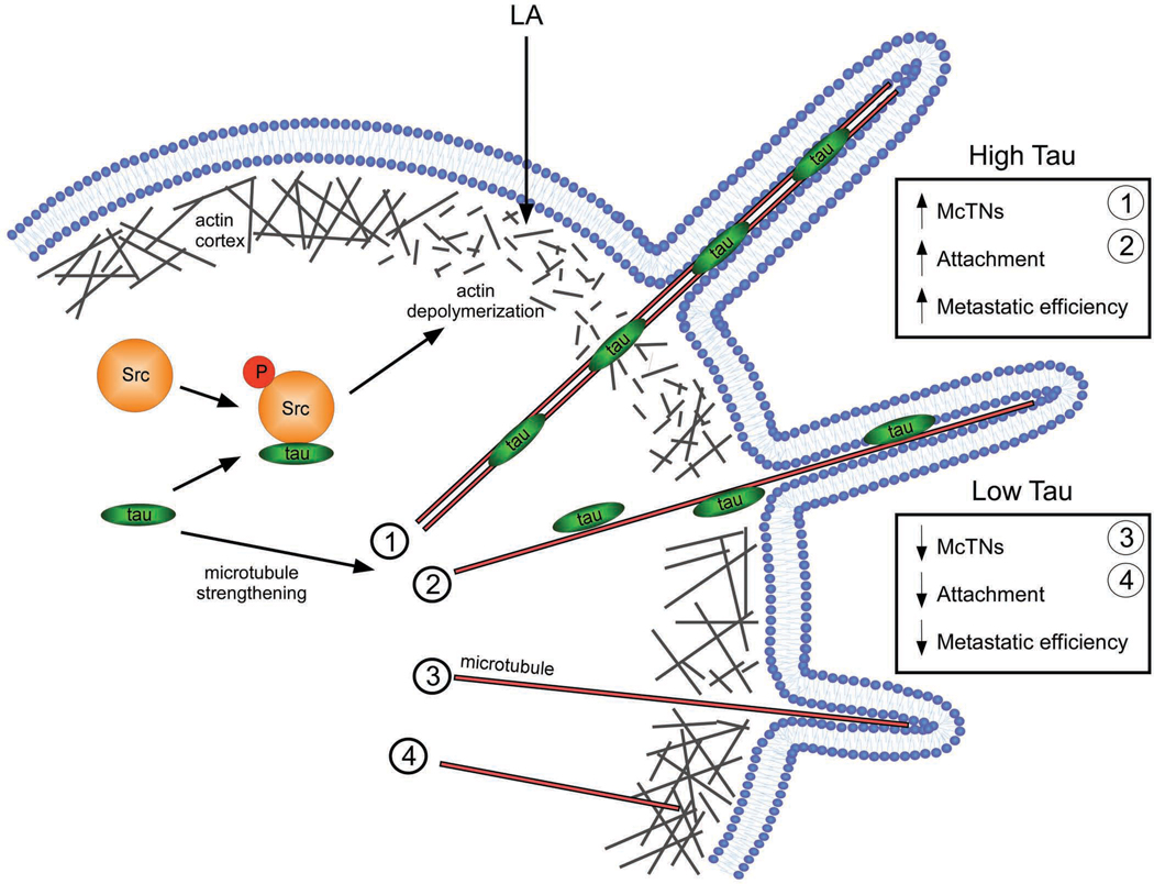
Figure 6. Model of tau-induced McTN formation.
In detached CTCs expressing tau, tau binds to and stabilizes microtubules, inducing microtubule bundling [1] and/or increasing individual polymer strength [2]. These microtubules are more able to penetrate the actin cortex due to preexisting defects in actin bundling or tau-induced actin depolymerization via priming of Src phosphorylation (Sharma et al., 2007). Consequently, microtubules easily deform the plasma membrane into McTNs that increase the ability of the cell to reattach to an ECM and improve metastatic efficiency. In detached CTCs lacking tau, microtubules are more dynamic and weaker; therefore, they are only able to form short McTNs [3] on none at all [4] due to the constriction of the actin cortex, even in the presence of preexisting cortical weaknesses. The lack of tau expression results in decreased efficiency of attachment and metastasis.

