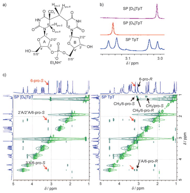
Figure 1.
a) Structure of isolated SP [D4]TpT. b) Zoom-in view of the 1H NMR signals for the 5′-T 6-Hpro-S and 6-Hpro-R hydrogen atoms in SP [D3]TpT, SP [D4]TpT, and SP TpT. c) Zoom-in view of the ROESY spectra of SP TpT and SP [D4]TpT dissolved in [D6]dimethyl sulfoxide. The signals associated with 6-Hpro-S are indicated by red arrows; these signals are observed for both SP species. The signals associated with 6-Hpro-R or CH3 are indicated by black arrows; these signals are not present in the spectrum for SP [D4]TpT as a result of the deuterium substitution. The interaction between the 5′-T 6-Hpro-S and 3′-T 6-H atoms yields a ROESY signal at δ=3.14–7.51 ppm, which is outside the range of the zoom-in view. The full 1D and 2D NMR spectra for these SP species can be found in the Supporting Information.

