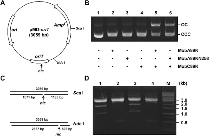
Fig. 5.

Site- and strand-specific relaxation of the 89K oriT region.
A. Physical map of the pMD-oriT plasmid, which contains the oriT region of 89K and serves as substrate DNA in the relaxation analysis. The putative oriT nick site (nic) is indicated by a vertical arrow.
B. Equivalent substrate pMD-oriT plasmid was incubated with (+) or without (–) the proteins of interest. Reaction products were analysed by standard agarose gel electrophoresis (0.9%). OC, open circular plasmid DNA; CCC, covalently closed circular plasmid DNA.
C. Schematic representation of the expected single-strand species generated by a site-specific nick (arrow) on endonuclease-linearized pMD-oriT DNA. The size of each ssDNA fragment is shown.
D. pMD-oriT was linearized with either ScaI or NdeI, and the relaxation mixtures were analysed on a 0.9% alkaline agarose gel. Lanes: 1, ScaI-linearized pMD-oriT with MobA89K and MobC89K; 2, NdeI-linearized pMD-oriT with MobA89K and MobC89K; 3, ScaI-linearized pMD-oriT with MobA89KN258 and MobC89K; 4, NdeI-linearized pMD-oriT with MobA89K and MobC89K. The 1 kb DNA ladder marker is shown to the right (M).
