
Figure 4.
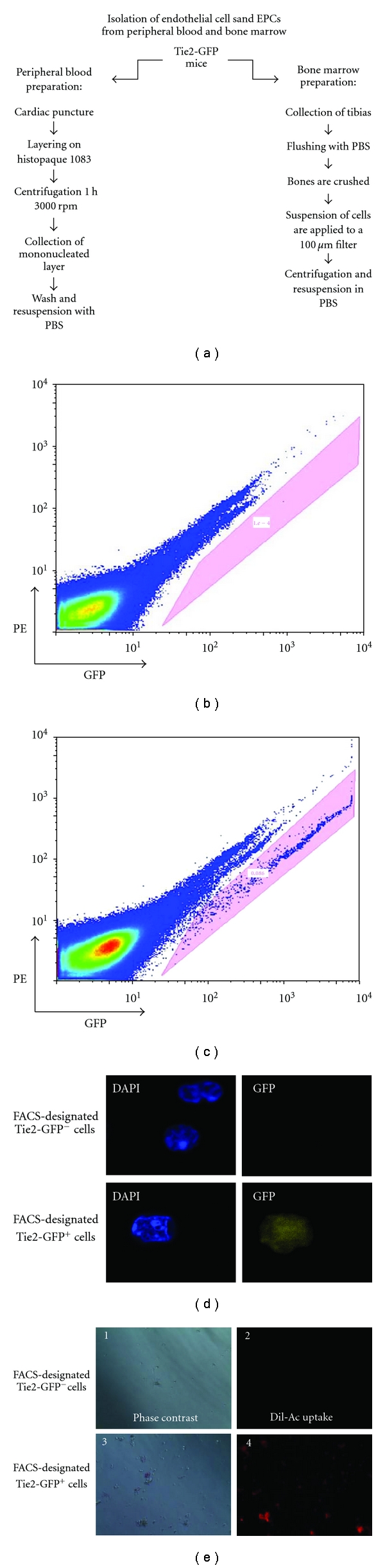
Isolation of Tie2-GFP+ cells from bone marrow and peripheral blood in mice. (a) Isolation of bone marrow and peripheral blood cells was performed in parallel as depicted. Washed cells were resuspended in ice-cold PBS and shielded from the light to prevent photobleaching. (b) and (c) Flow cytometric analysis of isolated cells. Resuspended cells were subjected to FACS on an Influx cell sorter. The profile obtained from Tie2-GFP mice (c) exhibited an additional population when compared to the wild-type C57BL/6 (b) (denoted in the shaded pink box). Therefore, this exclusive population was characteristic of the Tie2-GFP+ cells. (d) Tie2-GFP− and Tie2-GFP+ cells as identified by FACS analysis were collected separately, fixed onto slides, counter-stained with DAPI, and visualized by fluorescence microscopy. Only cells identified by prior FACS analysis as Tie2-GFP+ exhibited significant levels of green fluorescence by microscopic (100x) analysis. (e) Tie2-GFP+ cells which were isolated by FACS from Tie2-GFP mice were viable and of the endothelial cell lineage, as demonstrated by their ability to uptake Dil-Ac-LDL. Cells sorted by FACS as Tie2-GFP− did not have the ability to uptake Dil-Ac-LDL.