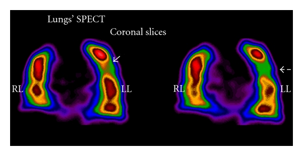
Figure 11.

Two sequential coronal slices indicating mild pulmonary embolism in left lung lobe (LL) not indicating in planar images. Image reconstruction has been completed by FBP and Butterworth filter (critical frequency 0.5, order 10). Courtesy of M. Gavrilelli (MSc, “Medical Imaging Center” Athens, Greece).
