Figure 3.
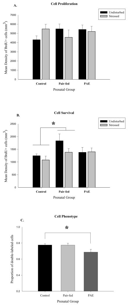
A. Density of BrdU-ir cells in the granule cell layer (GCL) 24 hours after BrdU administration (n = 4-7 per group). No significant differences in cell proliferation among groups. B. Density of BrdU-ir labeled cells measured 3 weeks after BrdU administration (n = 4-8 per group). Within the GCL, Pair-fed > Control (p = 0.03). C. Phenotype of surviving cells (n = 50 cells/rat). The proportion of double-labeled (NeuN/BrdU or GFAP/BrdU) cells, collapsed across stress condition, was reduced in rats prenatally exposed to alcohol (PAE < Control (p = 0.02)).

