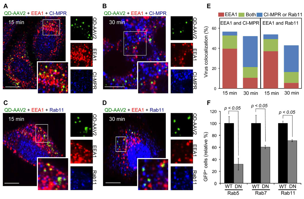
Figure 4.
The trafficking of QD-AAV2 through various endosomes. (A to D) HeLa cells were incubated with QD-AAV2 (green) for 30 min at 4°C to synchronize infection and were then shift to 37°C for 15 or 30 min. The cells were fixed, permeabilized, and immunostained with antibodies against EEA1 (red) and CI-MPR (blue) (A, B), or EEA1 and Rab11 (blue) (C, D). The boxed regions are enlarged in the right panels. Scale bar represents 5 µm. (E) Quantification of QD-AAV2 colocalized with EEA1+, CI-MPR+, Rab11+, EEA1+CI-MPR+, or EEA1+Rab11+ endosomes after 15 or 30 min of incubation. (F) Functional involvement of endosomes in the AAV2 transduction. 293T cells transiently transfected with the wild-type or dominant-negative mutant form of Rab5, Rab7, or Rab11 were infected with unlabeled AAV2. The percentage of GFP-positive cells was analyzed by flow cytometry. Error bars represent the standard deviation of the mean from triplicate experiments.

