Figure 5.
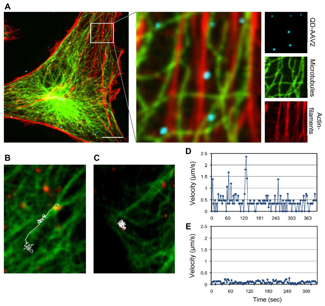
Cytoskeleton-mediated AAV2 transport. (A) QD-labeled AAV2 (blue) was incubated with HeLa cells for 30 min at 37°C. The cells were then fixed, permeabilized, and stained for microtubules (green) and actin-filaments (red) with the monoclonal antibody to α-tubulin and rhodamine-conjugated phalloidin, respectively. The boxed regions are enlarged in the right panels. (B to E) Live cell imaging of microtubule-dependent or microtubule-independent movement of QD-AAV2. HeLa cells were transiently transfected with GFP-α-tubulin. At 24 h post-transfection, the cells were incubated with QD-labeled AAV2 (green) for 30 min at 4°C, and were then warmed to 37°C for 15 min. Confocal time-lapse images were then recorded. A representative trajectory and viral velocity of microtubule-dependent (B, D) and microtubule-independent movement (C, E) of QD-AAV2 are shown. Scale bars represent 5 µm.

