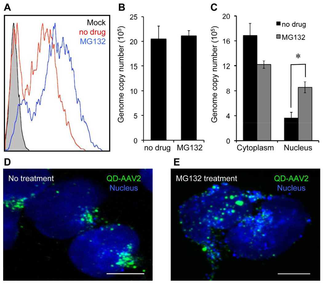
Figure 6.
Enhanced nuclear uptake of AAV in the presence of the proteasome inhibitor. (A) HeLa cells were pretreated with MG132 (25 µM) for 30 min at 37°C and then spin-infected with AAV2 in the presence of MG132. After an additional 3 h of incubation with MG132 at 37°C, the cells were washed with PBS and resupplied with fresh media. The resulting GFP expression was analyzed by flow cytometry at 3 days post-infection. (B) Quantification of the intracellular viral genome copy number in infected cells with or without MG132 at 24 h post-infection. (C) Quantification of intracellular viral genomes isolated from the cytoplasmic and nuclear fractions in infected cells with or without MG132 at 24 h post-infection. Error bars represent the standard deviation of the mean from triplicate experiments (* p<0.05). (D, E) Enhanced nuclear translocation of QD-AAV2. HeLa cells were incubated with QD-AAV2 (green) for 18 h at 37°C with (E) or without MG132 (D). The cells were then fixed, permeabilized, and counterstained with TO-PRO-3 (blue). Scale bars represent 5 µm.

