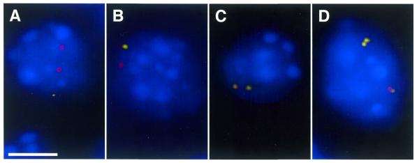
Figure 1.
RNA–DNA FISH examples. Pax6 RNA–DNA FISH performed on E12.5 neural retina cells reveals DAPI-stained nuclei (blue) with no (A), one (B), two (C) or four (D) spots of Pax6 RNA (green) co-localizing to either two (A–C) (G1 cells) or four (D) (G2 cells) Pax6 DNA alleles (red). The scale bar in (A) represents 5 µm.

