
Figure 3.
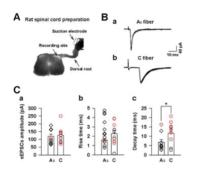
GRP sensitive spinal dorsal horn neurons mostly receives sensory inputs from C fibers. (A) Digitized photomicrograph showing one example for whole cell patch recording on neurons in the superficial lamina of spinal cord, which was stimulated by dorsal roots. (B) Examples of Aδ (a) and C fiber (b) evoked monosynaptic EPSCs. (C) The amplitude (a), rise time (b) and decay time (c) of Aδ and C fiber evoked EPSCs. The Red and black showed GRP sensitive and insensitive neurons, respectively. * P < 0.05, significant difference between Aδ and C fiber.