
Figure 6.
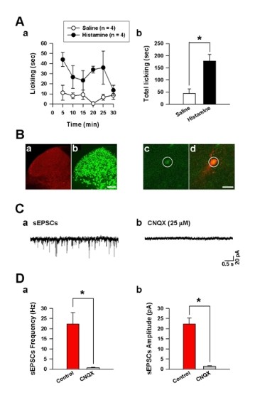
Identification of the transmitter on FosGFP-expressing neurons in the spinal cord after intradermal histamine injection in FosGFP transgenic mice. (A) (a) Time course of intradermal histamine (black circles, n = 4) and saline (white circles, n = 4) induced licking behaviors every 5 min for 30 min in FosGFP transgenic mice. (b) The intradermal histamine injection significantly increased total licking behaviors for 30 min compared to the intradermal saline injection. (B) (a) Fos-positive cells were found in the superficial dorsal horn of adult mice 120 min after histamine injection. (b) Dual immunostaining of NeuN in the dorsal horn was also shown. Scale bar, 100 μm. Patch clamp recordings were shown from FosGFP-expressing neurons in the spinal cord. Images showed that one of the FosGFP-expressing neurons (c) in the spinal cord was recorded and labeled by Alexa fluor 594 (d). Yellow color indicated the overlap of GFP and Alexa fluor 594 (d). Scale bar, 20 μm. (C) Spontaneous EPSCs (sEPSCs) recorded on the FosGFP-expressing neurons of the dorsal horn (a) were completely blocked by a bath application of CNQX (25 μM) (b). (D) The summarized data showing that the frequency (a) and amplitude (b) of sEPSCs in histamine induced FosGFP-expressing neurons were totally blocked by CNQX (n = 4).