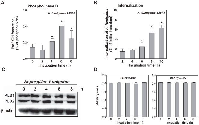
Figure 1. A. fumigatus stimulates PLD activity during its internalization into A549 cells.
A. A549 cells were prelabeled with [3H] oleic acid and infected with the resting conidia of A. fumigatus 13073 at an MOI of 10 for the indicated time periods. Then, ethanol was added to determine the PLD activity. B. A549 cells were infected with the resting conidia of A. fumigatus 13073 at an MOI of 10 for the indicated time periods, and the internalization of A. fumigatus was analyzed by the nystatin protection assay. Differences in [3H] PtdEtOH formation between the 0 h time point and the other time points (A) and differences in the internalization of A. fumigatus between the 2 h time point and the other time points (B) were compared. In parallel, the cells were lysed for immunoblotting with the indicated antibody (C) and the densitometric analysis of immunoblots for three independent experiments is shown (D). Data are represented as the mean ± SE (n = 3–4), and the blots are characteristic of 3 independent experiments. *P<0.05.

