
Figure 1.
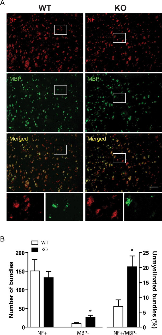
PGC1α KO mice displayed developmental myelination deficit in the striatum. A, Coronal brain sections from P10 PGC1α KO and WT mice (n = 5 each) were immunostained with NF (red) and MBP (green) antibodies. B, While there was no significant difference in the total number of NF+, the number of NF+/MBP− and the percentage of NF+/MBP− were significantly increased in PGC1α KO mice as compared with littermate WT controls. Data expressed as mean ± SEM. Student's t test; *p < 0.05. Scale bar, 100 μm.