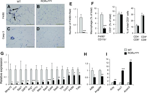
Fig. 7.
WAT-associated inflammatory response is significantly reduced in WAT of RORαsg/sg(HFD) mice. A, B: macrophage infiltration into eWAT was greatly reduced in RORαsg/sg(HFD) mice. F4/80+ macrophages were identified by immunohistochemical staining as “crown-like structures” (arrows). Scale bar indicates 200 μm. C, D: reduced apoptosis in eWAT of RORαsg/sg(HFD) mice compared with WT(HDF). Apoptotic cells were stained with an antibody for active-caspase 3. E: the number of F4/80-positive cells were decreased in WAT of RORαsg/sg(HFD) than WT(HFD) mice (WT, n = 5; RORαsg/sg, n = 5). F: stromal-vascular fraction cells from eWAT of WT(HFD) and RORαsg/sg(HFD) were examined by flow cytometry analysis as described in materials and methods. The percentage of the macrophage population (F4/80+CD11b+ cells) was significantly reduced, but T cell population (CD3+ cells) and CD4+ and CD8+ subpopulations were unaffected in RORαsg/sg(HFD) mice (WT, n = 9; RORαsg/sg, n = 5). G, H: induction of inflammatory genes and metabolic in WAT was greatly decreased in RORαsg/sg(HFD) for 6 wk. I: induction of lipogenic genes in WAT of RORαsg/sg(HFD) mice. Gene expression was analyzed by QRT-PCR (5 male mice in each group). Data present means ± SE. *P < 0.05, **P < 0.01, ***P < 0.001.

