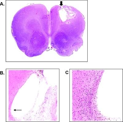
Figure 7.
Histologic analysis of coronal sections of brains of tumor bearing rats after treatment with 40 mM ACCA through CED. (A) Representative H&E-stained coronal section (x10) of brain of one of the animals that survived beyond experimental end points. Cannulation/ posttumor necrotic tissue cavity (striatal region; arrow) 120 days after CED of ACCA. (B) Necrotic cavity (arrow) at x40 magnification. (C) Cavity wall at x200 magnification.

