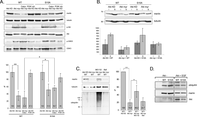
Figure 3.
Constitutively active Akt degrades WT merlin through the proteasomal pathway. (A) COS-7 cells cotransfected with WT or S10A merlin and inactive (Akt KD) or constitutively active (Akt myr) Akt were untreated or treated with the phosphatase inhibitor Calyculin A (Calyc.) or PI3K inhibitor LY294002 (PI3K inh.). Lysates were run in SDS-PAGE and detected for merlin (A-19 Ab), phospho-Ser 473 Akt (p-Akt), Akt, phospho-GSK3 (p-GSK3), GSK3, and α-tubulin as a loading control. The merlin/tubulin ratio is shown in the lower chart and 100% represents the merlin level in Akt KD-expressing cells. The amount of WT merlin in cells expressing Akt myr is significantly decreased compared with cells expressing Akt KD, whereas S10A merlin is less sensitive to Akt myr coexpression. (B) Lysates from COS-7 cells cotransfected with merlin and Akt KD or Akt myr were separated into insoluble (I) and soluble (S) fractions and run in SDS-PAGE (upper panel). The merlin-tubulin ratio was quantified, and the amount of merlin in fractions is shown in arbitrary units (lower chart). Both the detergent-insoluble and -soluble WT merlin is degraded in Akt myr-transfected cells. (C) COS-7 cells were cotransfected with merlin WT and Akt KD or Akt myr. The WT + Akt myr-expressing cells were untreated or treated with MG132 or Bafilomycin A1 (Baf.), and lysates were detected with merlin A-19 Ab, α-tubulin Ab, and ubiquitin Ab (left panels). The merlin amount was quantified, and the level in WT + Akt KD-transfected cells was regarded as 100% (right chart). The proteasome inhibitor MG132 increases the amount of merlin in Akt myr-expressing cells. (D) WT and S10A merlin was immunoprecipitated from untransfected or Akt-transfected EGF-stimulated and MG132-treated COS-7 cells. Samples were detected for ubiquitin (upper panel) and merlin (middle panel). The expression of Akt was confirmed from the lysates with Akt antibody (lower panel). Merlin WT and S10A are equally ubiquitinated. **P < .01. *P < .05.

