Figure 1.
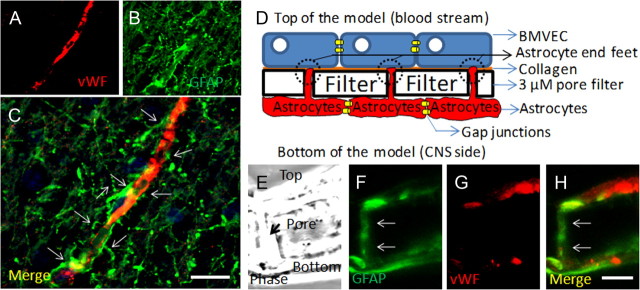
Astrocytes are key cells in the maintenance of the BBB in vivo and in vitro. A–C, Staining of human brain tissue sections for vWF (a marker of endothelial cells; red staining) (A) and GFAP (a marker of astrocytes; green staining) (B) demonstrates the close interaction between astrocytes and their end feet with endothelial cells in vivo (C) (merge of both colors). D, Schematic of our in vitro model of the human BBB, composed of BMVECs grown on denatured collagen on the top of a polycarbonate membrane with 3 μm pores, and human astrocytes cultured on the bottom side of the membrane. Astrocytes that are coupled by gap junctions send processes, termed astrocyte end feet, through the 3 μm pores. E–H, Staining of a cross section of the BBB model (see phase picture; E) for GFAP (F) or vWF (G) or the merge of both colors demonstrated that vWF staining is in the top of the insert and GFAP-positive astrocytes are on the bottom. Astrocytes send processes from the bottom of the insert to the top of the insert to establish contact with the BMVEC layer. Much of this GFAP staining is also detectable on the top of the insert (H; colocalization of both colors; yellow). Thus, our in vitro model of the human BBB recapitulates many of the astrocyte–endothelial interactions seen in vivo. This model will be used to examine the role of HIV-infected astrocytes in BBB integrity and astrocyte end feet signaling. Scale bar, 50 μm.

