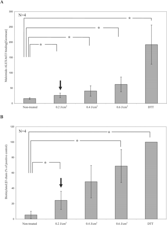
Fig. 5.
UVC light-induced reduction of disulfide bonds on the platelet surface. A The amount of free thiol groups on the platelet surface was measured by the help of maleimide which has a high binding affinity to free thiol groups. Platelets were either left untreated, irradiated with increasing doses of UVC light or treated with dithiothreitol (DTT) as positive control and then incubated with maleimide coupled to the fluorescent dye Alexa633 to label free thiol groups. Binding of the probe was assessed by flow cytometry. Data represent the mean of 4 experiments. B To investigate the effect on αIIbβ3 specifically, platelets were incubated with BMCC to biotinylate free thiol groups after the various treatments. Biotinylated proteins were then precipitated with streptavidin agarose beads. The graph depicts the binding of the β3 antibody to the immunoblots of the precipitates as quantified on an imaging system and calculated as percentage of the results of the positive control set as 100%. Data represent the mean of 4 experiments. Arrows indicate the dose of 0.2 J/cm2 as used in the THERAFLEX UV-Platelets system.
*Significant differences between negative controls and irradiated samples were detected at a p value of less than 0.05 (paired t-test).

