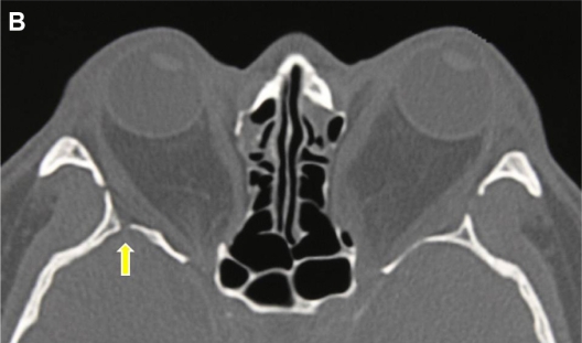
Figure 1B.
Axial computerized tomography scan of a 34-year-old male patient with Graves’ orbitopathy after bilateral balanced decompression surgery with rim intact approach. The deep lateral area of the trigone was not removed. The yellow arrow indicates the bony defect through which cerebrospinal fluid leakage occurred.

