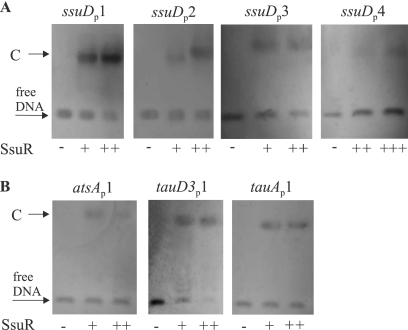
Fig. 5.
Binding of SsuR to short segments of four promoter regions. The EMSAs were performed on agarose gels with DNA probes (about 50 ng) containing portions of the ssuD, atsA, tauD3, and tauA promoter regions. Probes were preincubated with SsuR(His6) protein at a concentration of 5 μg/ml (+), 10 μg/ml (++), or 20 μg/ml (+++). Arrows indicate unbound probe (free DNA) and complexes (C) observed as shifted bands. (A) Four ssuDP probes containing sequences indicated as segments 1, 2, 3, and 4 in Fig. 4. (B) Probes designated atsAP1, tauD3P1, and tauAP1 contained segments indicated as sequence 1 (between > and <) in Fig. 4. No shifted band was produced by SsuR with a 50-bp fragment internal to ssuD ORF (data not shown).

