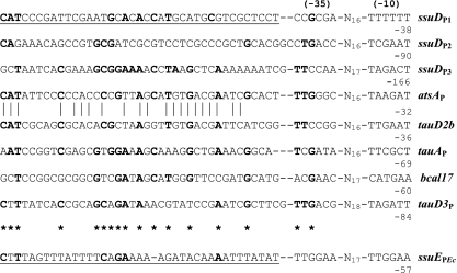
Fig. 6.
Alignment of identified and putative SsuR-binding sites at target promoter regions. Putative elements of σ70-dependent promoters in regions analyzed are indicated as (−35) and (−10). Coordinates relative to the translation start codons of downstream ORFs are indicated below the downstream limit of the −10 region in each case. The region of ssuDP protected by SsuR from DNase digestion (detectable at the lowest concentration of SsuR) is underlined. Nucleotides identical in at least five SsuR-regulated promoter regions are shown in bold and indicated by asterisks below the aligned sequences. The highest identity observed between atsAP and tauD2bP sequences is illustrated by vertical bars. For a comparison, the ssuE promoter region of E. coli (ssuEPEc) that is upregulated by CblEc (5) and SsuRBc (18) is also shown with the Cbl-binding site/putative SsuR-binding site underlined.

